
Vascular Remodeling of the Lumbosacral Enlargement
This illustration was produced with Dr. Phillipe Gailloud from the Department of Interventional Neuroradiology. It is intended to accompany a manuscript which will be submitted for publication.
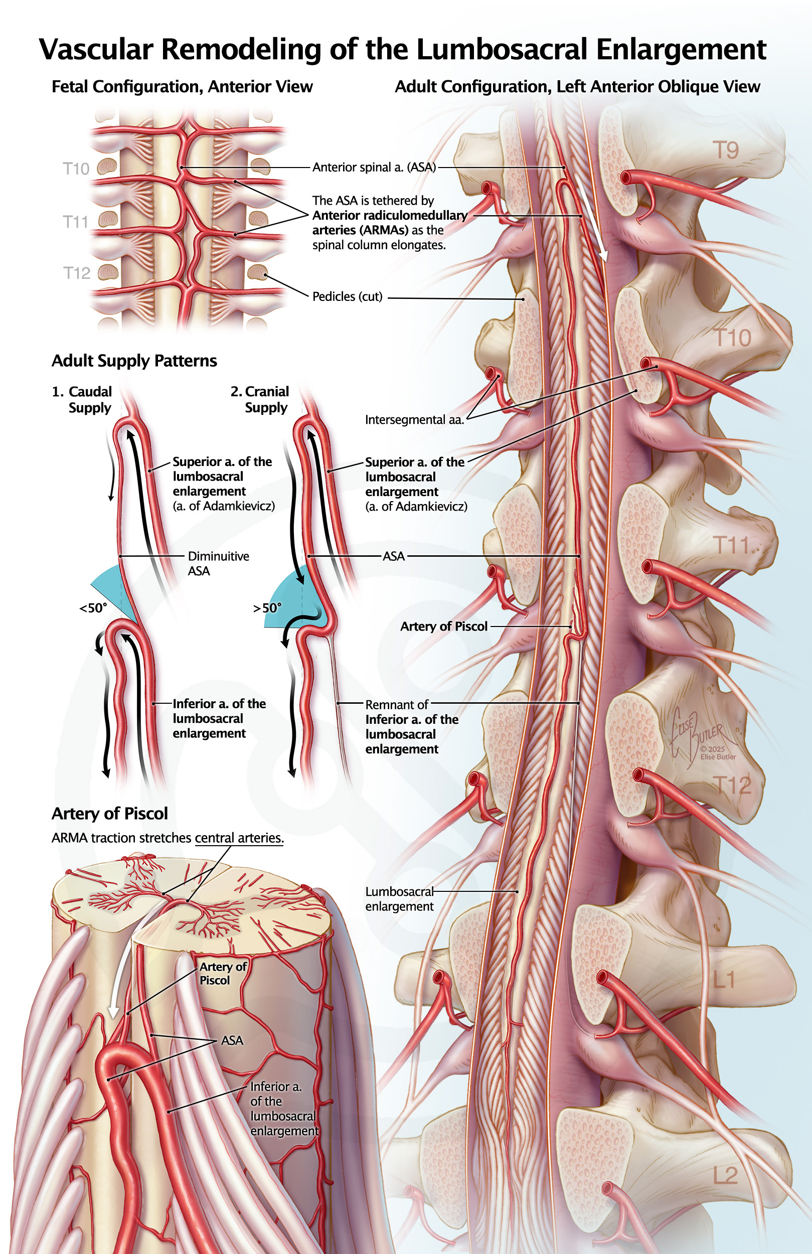
The upper left image depicts fetal anatomy, with anterior radiculomedullary arteries (ARMAs) traveling directly medial to supply the spinal cord. Shared labels connect this inset to the main view, showing how spinal column elongation creates traction on ARMAs, which can deflect the anterior spinal artery (ASA). A sagittal cut through the pedicles and dura provides a clear view. The middle left diagram demonstrates that if the angle of ASA deflection is less than fifty degrees, an ASA segment diminishes and the inferior ARMA (caudal supply) remains patent. If the angle of deflection exceeds fifty degrees, the cord receives a cranial supply and the inferior ARMA closes. Graphic elements clarify the angles of deflection, and black arrows describe the hemodynamics of the ASA. The bottom inset shows how ARMA traction on the ASA can stretch central arteries, creating an Artery of Piscol.
The upper left image depicts fetal anatomy, with anterior radiculomedullary arteries (ARMAs) traveling directly medial to supply the spinal cord. Shared labels connect this inset to the main view, showing how spinal column elongation creates traction on ARMAs, which can deflect the anterior spinal artery (ASA). A sagittal cut through the pedicles and dura provides a clear view. The middle left diagram demonstrates that if the angle of ASA deflection is less than fifty degrees, an ASA segment diminishes and the inferior ARMA (caudal supply) remains patent. If the angle of deflection exceeds fifty degrees, the cord receives a cranial supply and the inferior ARMA closes. Graphic elements clarify the angles of deflection, and black arrows describe the hemodynamics of the ASA. The bottom inset shows how ARMA traction on the ASA can stretch central arteries, creating an Artery of Piscol.

AMI Salon 2026
Student Best in Show
AMI Salon 2026
Award of Excellence