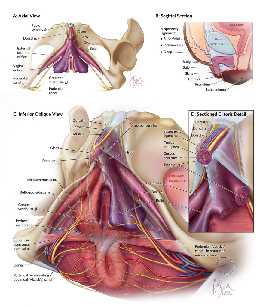

Anatomy of the Clitoris
This illustration, produced with content experts from the Johns Hopkins Department of Radiology and Center for Functional Anatomy and Evolution, depicts the anatomy of the clitoris and surrounding structures. It is intended for a broad audience, but we chose the orienting views for their familiarity to radiologists.
This project was conceived due to the dearth of research and depictions of the clitoris in medical literature, despite its relevance in patient care. The piece is published as a full-page spread as part of an article in the Journal of Abdominal Radiology.
Please check out the case study below to see what went into creating this piece!
Dissections
I observed two dissections of the perineum and clitoris which were performed by Dr. Siobhán Cooke from the Center for Functional Anatomy and Evolution. These are some quick sketches created from observation of the cadavers. I was also permitted to take photos.
I previously had the opportunity to dissect the female perineum in our anatomy class, directed by Dr. Cooke.
Radiological Data
I viewed radiographs from several individuals using HOROS, an open-source medical image viewer. Based on personal observation, the position and size of the clitoris vary somewhat between individuals.
Using 3D Slicer, I performed a very rough segmentation of one radiograph (seen to the right) to establish the relationship of the clitoris to the surrounding anatomy, as well as its 3D form.
The clitoris is a soft tissue structure which blends into the surrounding tissue on imaging, and in some areas my segmentation overreached its actual boundaries. I supplemented my segmentation by viewing 3D reconstructions from the literature.

Literature Review
I referred to previous literature reviews, and performed my own, to gather as much information about clitoral anatomy (as we currently understand it) as I could. My sources are cited to the right.
The anatomy of the clitoris is an area of active study, and our understanding is still evolving.
Content Expert Review
In addition to my own research, I was advised and educated by my content experts, Dr. Erin Gomez and Dr. Siobhan Cooke, and my teacher, Lydia Gregg.
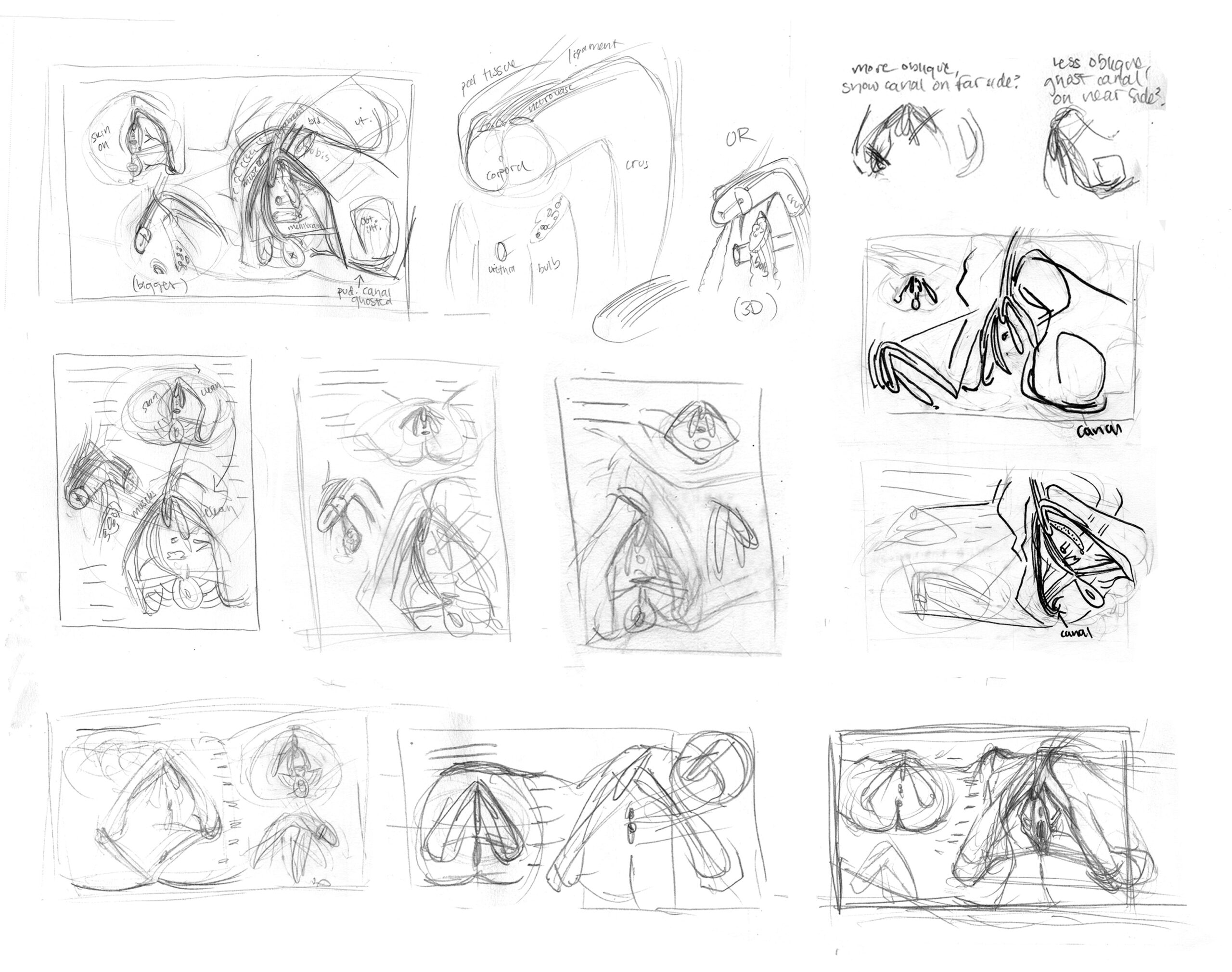
Thumbnails
I created small thumbnail illustrations to plan the content and layout of the piece. We knew we wanted a dynamic view which emphasized the 3D structure of the clitoris, that the pudendal canal should be indicated, and that an axial view should be included for radiologists.
The elements and layout of the piece evolved throughout the process. A selection of initial ideas are shown to the right.
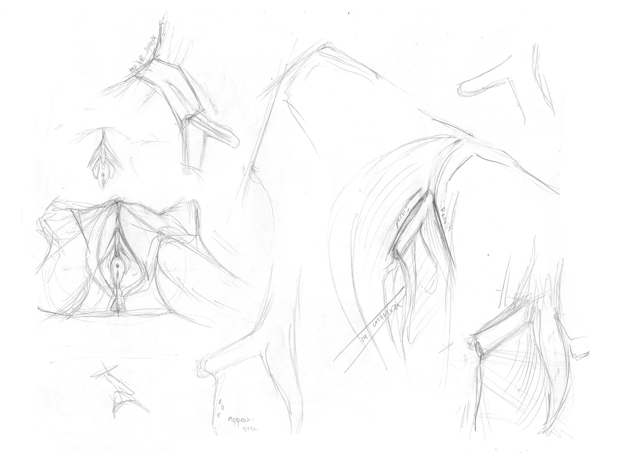
Neurovasculature
To make sure I understood the nerves, arteries, and veins in this area, I referred to multiple sources. Grey’s Anatomy contains detailed descriptions of the courses and destinations of the perineal vessels and nerves. I created shorthand sketches and notes to remind myself where to place them, even though they would not be labeled in the finished piece.
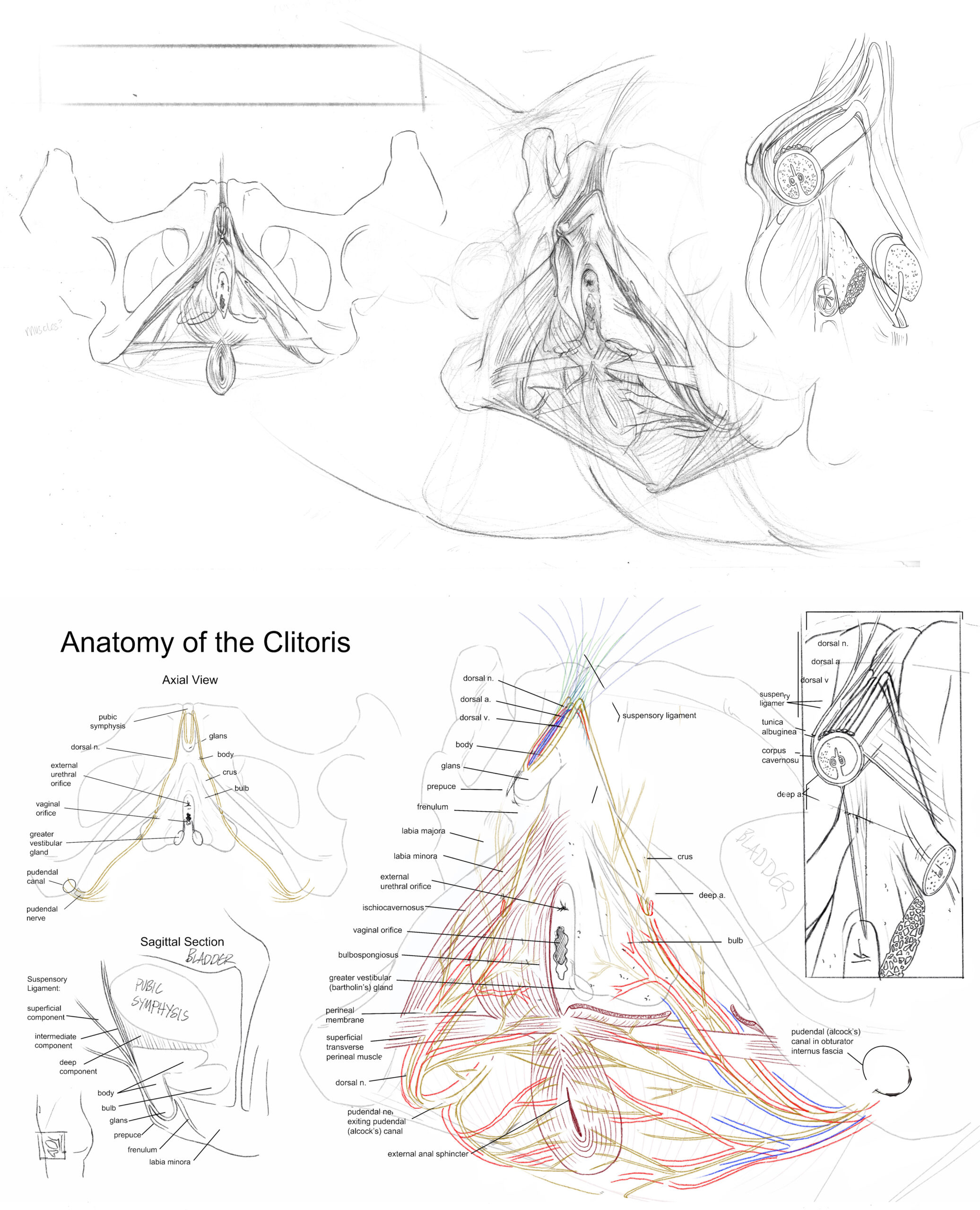
Rough Sketches
At each checkpoint in the process, I checked in with my content experts to ensure the illustrations were accurate and were achieving their goals. To the upper right are my first rough sketches. Below is a rough layout created after receiving feedback, once we had decided to add a sagittal view. I made an effort to clarify the structures in the second layout.
My goal in these early stages was not to create something beautiful, but to establish a foundation which was accurate and didactically successful.
Publication
This piece was not created for publication, but I was approached by Drs. Gomez and Cooke with the request to include it in the paper: “Under the Hood: Vulvar Anatomy and Pathology With a Focus on MRI.”
I reformatted the piece from an anatomical plate to a full-page color layout for a journal, including labels for a figure legend.
The open-access article may be found here:
Golden E., Cooke S., Butler E., Gregg L. & Gomez, E. Under the hood: vulvar anatomy and pathology with a focus on MRI. Abdom Radiol (2025). https://doi.org/10.1007/s00261-025-05179-1